Home

Hören Wanne verdächtig gradient echo mri brain Herbst Stroh Haiku

Diagnostic value of T2*-weighted gradient-echo MRI for segmental evaluation  in cerebral venous sinus thrombosis. | Semantic Scholar
Diagnostic value of T2*-weighted gradient-echo MRI for segmental evaluation in cerebral venous sinus thrombosis. | Semantic Scholar

MRI of brain T2-weighted axial gradient echo sequence. a At diagnosis.... |  Download Scientific Diagram
MRI of brain T2-weighted axial gradient echo sequence. a At diagnosis.... | Download Scientific Diagram

Isolated foot drop due to subcortical hemorrhage | SpringerLink

Automated detection of cerebral microbleeds on T2*-weighted MRI |  Scientific Reports
Automated detection of cerebral microbleeds on T2*-weighted MRI | Scientific Reports

Gradient echo sequences | Radiology Reference Article | Radiopaedia.org

Susceptibility weighted imaging - Wikipedia

Gradient echo imaging - Markl - 2012 - Journal of Magnetic Resonance Imaging  - Wiley Online Library
Gradient echo imaging - Markl - 2012 - Journal of Magnetic Resonance Imaging - Wiley Online Library

Improved T2* Imaging without Increase in Scan Time: SWI Processing of 2D Gradient  Echo | American Journal of Neuroradiology
Improved T2* Imaging without Increase in Scan Time: SWI Processing of 2D Gradient Echo | American Journal of Neuroradiology
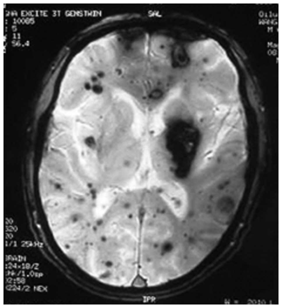
The value of T2*-weighted gradient echo imaging for detection of familial  cerebral cavernous malformation: A study of two families
The value of T2*-weighted gradient echo imaging for detection of familial cerebral cavernous malformation: A study of two families

Spoiled GRE - Questions and Answers ​in MRI

Importance of T2*-weighted gradient-echo MRI for diagnosis of cortical vein  thrombosis - European Journal of Radiology
Importance of T2*-weighted gradient-echo MRI for diagnosis of cortical vein thrombosis - European Journal of Radiology

T1, T2, FLAIR, and Gradient Echo pulse sequences. - YouTube

Brain MRI. Flair sequence (a) and gradient echo sequence(b). Initial... |  Download Scientific Diagram
Brain MRI. Flair sequence (a) and gradient echo sequence(b). Initial... | Download Scientific Diagram

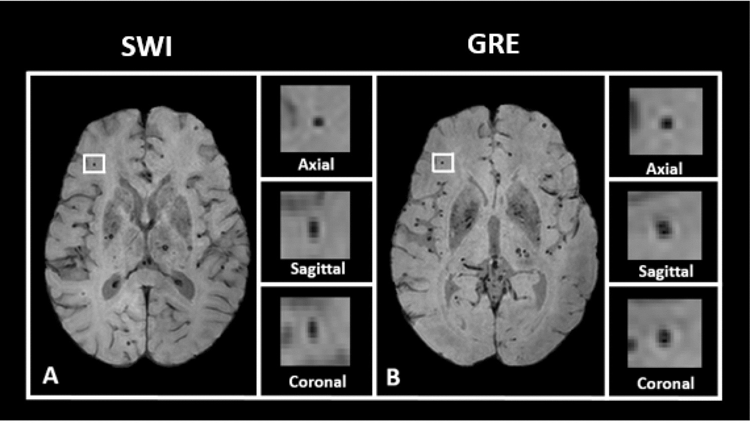
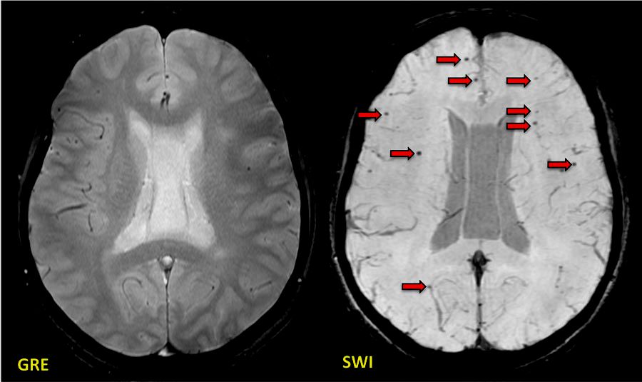
Superiority of SWI over GRE - Questions and Answers ​in MRI

STrategically Acquired Gradient Echo (STAGE) imaging, part I: Creating  enhanced T1 contrast and standardized susceptibility weighted imaging and  quantitative susceptibility mapping - SpinTech - MRI Technologies
STrategically Acquired Gradient Echo (STAGE) imaging, part I: Creating enhanced T1 contrast and standardized susceptibility weighted imaging and quantitative susceptibility mapping - SpinTech - MRI Technologies

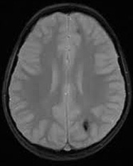
MRI brain (Gradient echo images) showed acute hemorrhages... | Download  Scientific Diagram
MRI brain (Gradient echo images) showed acute hemorrhages... | Download Scientific Diagram

Fig 1. | Brain Microhemorrhages Detected on T2*-Weighted Gradient-Echo MR  Images | American Journal of Neuroradiology
Fig 1. | Brain Microhemorrhages Detected on T2*-Weighted Gradient-Echo MR Images | American Journal of Neuroradiology
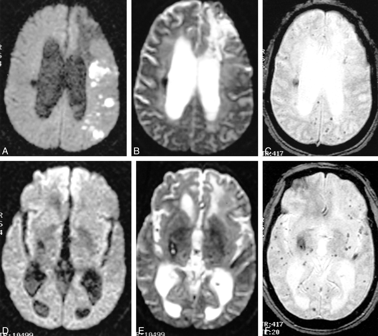
Detection of Intracranial Hemorrhage: Comparison between Gradient-echo  Images and b0 Images Obtained from Diffusion-weighted Echo-planar Sequences  | American Journal of Neuroradiology
Detection of Intracranial Hemorrhage: Comparison between Gradient-echo Images and b0 Images Obtained from Diffusion-weighted Echo-planar Sequences | American Journal of Neuroradiology

The value of T2*-weighted gradient-echo MRI for the diagnosis of cerebral  venous sinus thrombosis - Clinical Imaging
The value of T2*-weighted gradient-echo MRI for the diagnosis of cerebral venous sinus thrombosis - Clinical Imaging

Detection of Intracranial Hemorrhage: Comparison between Gradient-echo  Images and b0 Images Obtained from Diffusion-weighted Echo-planar Sequences  | American Journal of Neuroradiology
Detection of Intracranial Hemorrhage: Comparison between Gradient-echo Images and b0 Images Obtained from Diffusion-weighted Echo-planar Sequences | American Journal of Neuroradiology

MRI Protocols: Gradient echo sequence use in MRI BRAIN

Gradient echo MRI | Neurology

MRI BRAIN with T1 T2 (top left and right) and gradient echo (GRE,... |  Download Scientific Diagram
MRI BRAIN with T1 T2 (top left and right) and gradient echo (GRE,... | Download Scientific Diagram

GRE v SE - Questions and Answers ​in MRI

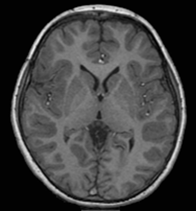
Gradient echo T1 (left: TR, 311 ms; TE, 2.5 ms; section thickness, 5... |  Download Scientific Diagram
Gradient echo T1 (left: TR, 311 ms; TE, 2.5 ms; section thickness, 5... | Download Scientific Diagram